Fig. 3.

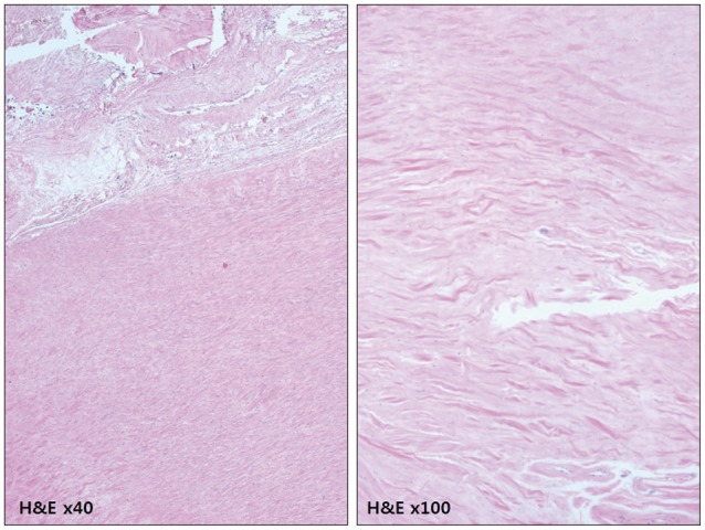
Photomicrograph of a section of the excised dura mater showed fibrotic pachymeninges with no significant inflammation, which was more apparent at the higher magnification view (Hematoxylin and eosin staining).

Photomicrograph of a section of the excised dura mater showed fibrotic pachymeninges with no significant inflammation, which was more apparent at the higher magnification view (Hematoxylin and eosin staining).