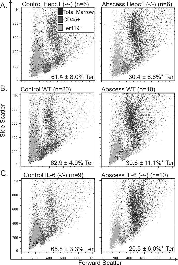
Figure 2.
Frequency of bone marrow erythroid progenitors and precursors in mice with sterile abscesses. We performed immunophenotyping of the marrow erythroid compartment. Orange represents Ter119-positive (Ter+) cells. Blue represent CD45-positive cells. Black represents cells that did not stain with either marker. The percentage of erythroid progenitors and precursors decreased from (A) 61.4 ±8.0% in untreated HepC1 (−/−) mice (N = 6) to 30.4 ± 6.6% in HepC1 (−/−) mice with sterile abscesses (N = 6); (B) from 62.9 ± 4.9 in untreated wild-type (WT) mice (N = 20) to 30.6 ± 11.1% in WT mice with sterile abscesses (N = 10); (C) from 65.8 ±3.3% in untreated IL-6 (−/−) mice (N = 9) to 20.5 ± 6.0% in IL-6 (−/−) mice with sterile abscesses (N = 10). Consistent with the increase in circulating neutrophils, granulocytes (CD45-positive marrow cells with high forward and high side scatter) expanded in mice with sterile abscesses (A–C). Asterisk indicates P <0.05. [Color figure can be viewed in the online issue, which is available at wileyonlinelibrary.com.]

