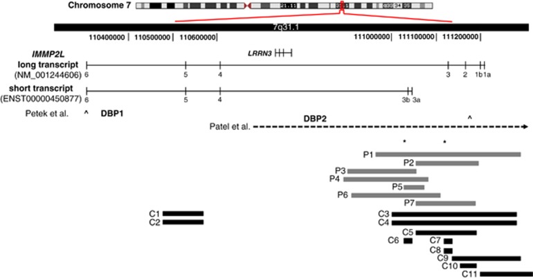
Figure 1.
Structural variations within the IMMP2L transcripts. (Genomic region showing the long (NM_001244606.1) and the short (ENST00000450877) transcripts of IMMP2L, and the structural variations identified in this study or by others. DBP1 and DBP2 indicate the duplication breakpoints and the open arrows indicate the amplicons (corresponding to exon 6 and exon 1b/IVS1a) used to screen for IMMP2L deletions (Petek et al7 and Petek et al,10 respectively). Horizontal dashed-arrow shows the 7.2-Mb deletion described by Patel et al.11 Horizontal gray blocks indicate the seven intragenic deletions (P1–P7) identified in Tourette patients in this study. The three deletions identified in the Danish background population (C6–C8) and the eight deletions identified in the Affymetrix CytoScan HD reference database (C1–C5 and C9–C11) are shown as horizontal black blocks. Asterisks represent localization of the qPCR amplicons.

