Figure 3.
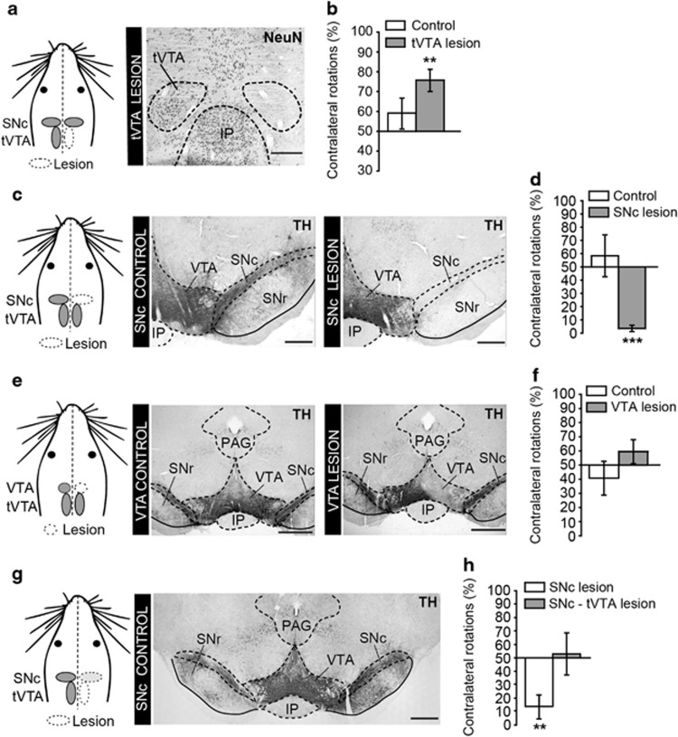
The tVTA controls rotation bias behavior. (a) Experimental protocol and histological evidence for unilateral tVTA lesion. (b) Unilateral tVTA lesion induces a contralateral rotation bias after amphetamine 3 mg/kg (control, n=17; lesion, n=12; **p<0.01). (c) Experimental protocol and histological evidence for 6-OHDA unilateral SNc lesion. (d) Unilateral SNc lesion induces an ipsilateral rotation bias after amphetamine administration (control, n=12; lesion, n=14; ***p<0.0001 referred to 50%). (e) Experimental protocol and histological evidence for unilateral VTA lesion with ibotenic acid. (f) Unilateral VTA lesion does not induce a rotation bias after amphetamine administration (control, n=9; lesion, n=7; referred to 50%). (g) Experimental protocol for unilateral SNc–tVTA lesions. The microphotograph illustrates a left side partial SNc lesion. (h) A tVTA lesion prevents the amphetamine-induced ipsilateral rotation bias observed after partial unilateral SNc lesion (SNc lesion, n=8; SNc–tVTA lesion, n=7; **p<0.01 referred to 50%). Graphs represent mean±SEM. 6-OHDA, 6-hydroxydopamine; IP, interpeduncular nucleus; PAG, periaqueductal gray; SNc, substantia nigra pars compacta; SNr, substantia nigra pars reticulata; TH, tyrosine hydroxylase; VTA, ventral tegmental area. Scale bars, 400 μm (a), 500 μm (c, e, g).

