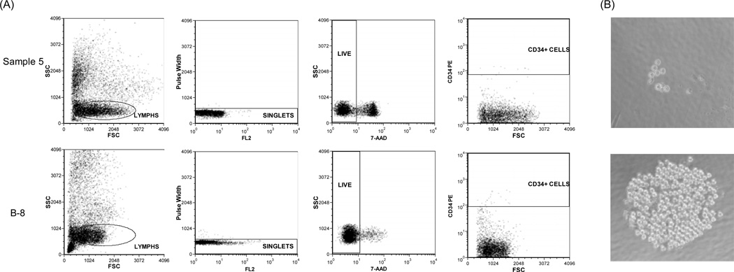
Figure 1.
Sorting for CD34+ cells using flow cytometry and culture assay for single CD34+ cells. (A) Dead cells in the mononuclear cells from donor #1 were demonstrated by staining with 7-AAD. Cells from Sample 5 (group I) had a lower level of viability than cells from B-8 (group II). (B) Single CD34+ cells from Sample 5 (group I) had less colony formation potential than those from B-5 (group II). A representative colony developed from individual CD34+ cell of each group was shown.

