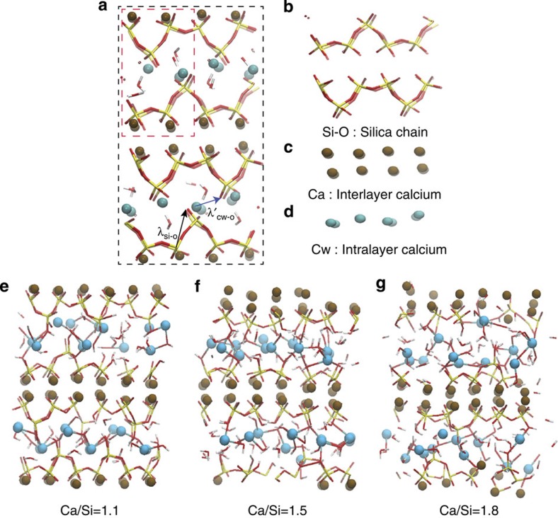
Figure 1. Effect of C/S ratio on the molecular structure of C-S-H at the nanoscale.
(a) The unit cell of tobermorite 11 Å is enclosed by black dashed line. The brown and cyan spheres represent intra- and interlayer calcium ions, respectively. Red and yellow sticks depict Si-O bonds in silicate tetrahedra. White and red sticks display hydroxyl groups and water molecules. By repetition of unit cells in all lattice vectors, a (2*2*2) supercell of the molecular structure of tobermorite 11 Å is constructed for representation and is outlined by dashed red line. The medium-range correlation lengths λ and λ’, which pertain to Si-O and Cw-O network are represented by dashed black and blue arrows, respectively. The solid skeleton of tobermorite consists of three parts: (b) silica chains, (c) calcium interlayer and (d) calcium intralayer. (e–g) Molecular model of C-S-H at C/S=1.1, 1.5 and 1.8. (e) At C/S=1.1, a lamellar structure is presented with minor defects in silica chains, reminiscent to that of 11 Å tobermorite. The interlayer regions contain counter charge-balancing calcium ions, hydroxyl groups and water molecules. (f) At C/S=1.5, several bridging tetrahedra are removed from the silicate chains. The interlayer calcium ions are still organized in well-defined sites. (g) The C/S ratio is further increased to 1.8 by removal of more silica tetrahedra. This indicates that from low to high C/S ratio, the C-S-H’s molecular structure changes from layered to a more amorphous structure.

