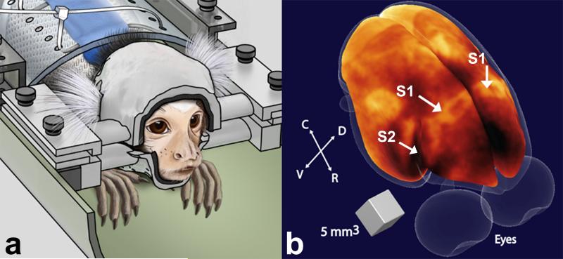
Figure 1.
(a) Illustration of a marmoset in the sphinx position restrained by the custom-fit helmet (left). (b) Volumetric rendering of the brain surface of a marmoset obtained from a 3D T1-weighted image (43). Regions of high myelination appear bright in the image. Arrows indicate the locations of primary (S1) and secondary (S2) somatosensory cortex. R = rostral; C = caudal; D = dorsal; V = ventral.

