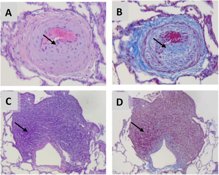
Figure 4.
Histologic findings in simian immunodeficiency virus–infected macaques are consistent with pulmonary hypertensive changes. Hematoxylin and eosin stain (A, C) and Masson trichrome stain (B, D). Arrows indicate medial hyperplasia and neointimal collagen deposition (A, B) and perivascular lymphoid tissue (C, D). Reprinted by permission from Reference 55.

