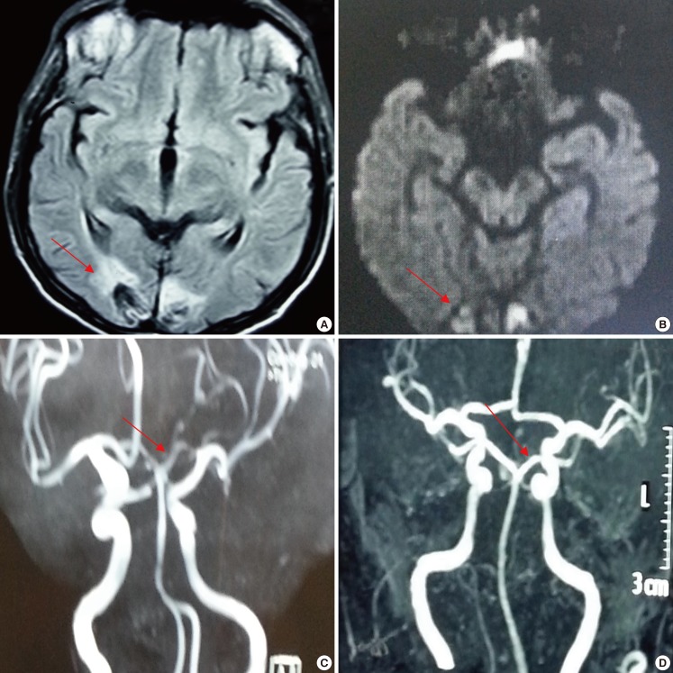
Figure 1.
Brain magnetic resonance imaging (MRI): (A) Fluid attenuated inversion recovery (FLAIR) sequence axial view showing hyperintensity in bilateral occipital lobe (red arrow); (B) Diffusion-weighted imaging (DWI) showing hyperintensity in bilateral occipital lobe (red arrow); (C) Brain magnetic resonance angiography (MRA) showing thinning of caliber of basilar artery and both posterior cerebral artery (PCA) (red arrow); (D) Repeated MRA after 3 months showing normalization of caliber of basilar artery and both PCA (red arrow).

