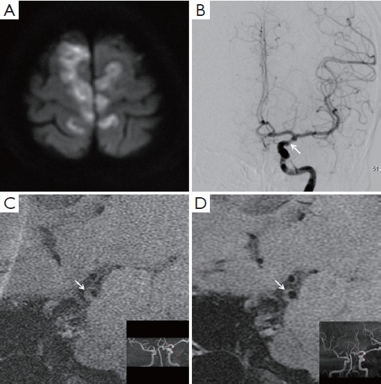
Figure 1.

Diffusion weighted imaging (A) and angiography (B) showed ischemic infarcts due to left internal carotid artery stenosis with an azygous anterior cerebral artery. On T1-weighted images of high-resolution MRI, a plaque (arrow, C) was identified, which was retracted (arrow, D; maximum plaque area from 0.15 to 0.10 cm2) after treatments.
