
Figure 1.
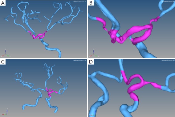
3D vascular models extracted from MRI images from two patients. The left column (A and C) is the main cerebral regions of interest arterial structure of the vascular surface. Please note the highlighted segments in purple (amplified on the right column, B and D).