
Figure 1.
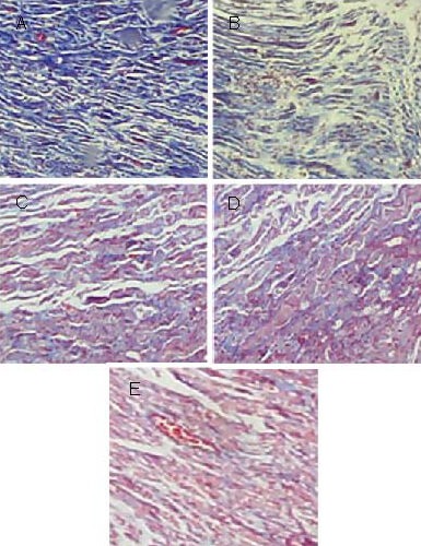
Collagen fiber proliferation in longitudinal sections of injured sciatic nerve at the nerve anastomosis site (Masson staining, light microscope, × 40).
In the model group (A), a large amount of collagen fibers were dyed dark blue.
In the tacrolimus gavage groups for 2 (B), 4 (C), 6 weeks (D), the number of blue collagen fibers gradually decreased, while of the number of red nuclei increased.
In the normal control group (E), the vast majority of staining was nuclear.