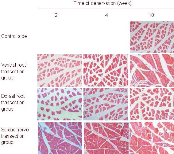
Figure 2.

Morphology of rat gastrocnemius muscle (hematoxylin-eosin staining, light microscope, × 100).
The size of gastrocnemius muscle cells gradually diminished in all rats after injury and reached the minimal value after 10 weeks in the sciatic nerve transection group.
The cell diameter and cross-section area were the largest in the dorsal root transection group.
