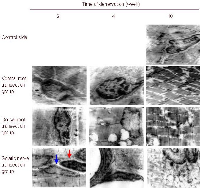
Figure 3.

Morphology of the rat gastrocnemius muscle (uranyl acetate-lead citrate staining, light microscope, × 8 000; blue arrow, muscle cell nuclei; red arrow, muscle satellite cell nuclei).
The ultrastructure of the gastrocnemius muscle cells exhibited similar changes in the three groups after nerve injury. With prolonged injury time, mitochondrial swelling, disorderly myofilaments and sarcomeres, shortened cristae, expanded sarcoplasmic reticulum, reduced glycogen granules, and enlarged nuclei were observed. In addition, myofilaments and sarcomeres broke, fused or disappeared, and mitochondria became vacuolated.
