Figure 3.

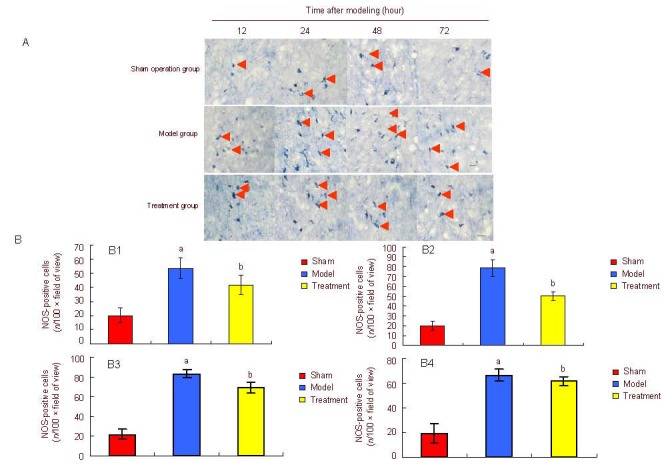
Effect of estrogen on nitric oxide synthase (NOS) expression in the medial septal nuclei of neonatal rats on the damaged side.
(A) Nicotinamide-adenine dinucleotide phosphate histochemical staining (bar: 100 μm) revealed the expression of NOS-positive neurons (arrows). NOS-positive cells in the model group showed small cell bodies and short neurites. The number of NOS-positive neurons in the treatment group was significantly lower than that in the model group.
(B) Quantification of NOS expression in medial septal nuclei of neonatal rats on the damaged side.
Data are expressed as mean ± SD of five rats in each group at each time point. At 12 (B1), 24 (B2), 48 (B3), 72 (B4) hours after modeling, aP < 0.05, vs. sham operation (sham) group; bP < 0.05, vs. model group, analysis of variance. Differences between two groups were compared using the least significant difference t-test.
