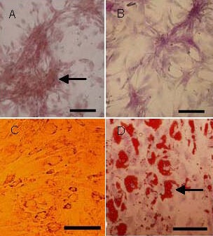
Figure 2.

Differentiation capacities of umbilical cord-derived mesenchymal stem cells (scale bars: 100 μm).
By the end of 14 days, osteogenic differentiation was reflected by the formation of a mineralized matrix (arrow) positively stained by alizarin red (A) and alkaline phosphatase expression (B). Adipocytic differentiation was demonstrated by the presence of a broadened morphology (C) and the formation of lipid vacuoles (arrow) by oil-red O staining (D).
