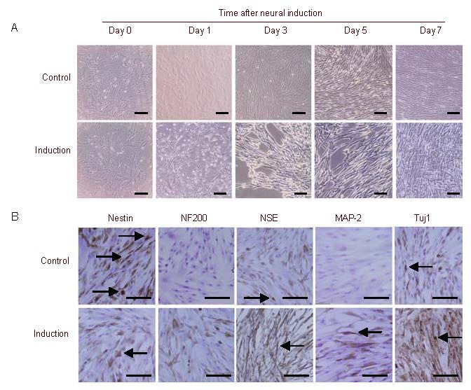
Figure 3.

Neural differentiation of umbilical cord-derived mesenchymal stem cells (UC-MSCs; scale bars: 100 μm).
(A) Morphological changes in MSCs following neural differentiation. Cells before induction were recorded as day 0. Cultures of untreated MSCs display a characteristic fibroblastic morphology. Neurally induced MSCs underwent morphological changes and acquired a pseudo-neuronal shape with neurite-like processes, whereas MSCs in the control group maintained their flattened morphology.
(B) Immunocytochemical analysis of neural marker expression by MSCs after neural differentiation. After 7 days of induction, cells expressed neurofilament 200 (NF200), neuron-specific enolase (NSE), microtubule-associated protein 2 (MAP-2) and Tuj1, while the untreated control group did not express these markers. Nestin expression was significantly reduced in the neural induction group. Arrows indicate positive expression.
