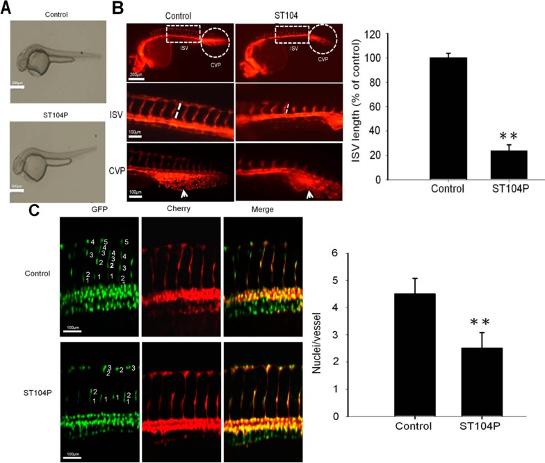
Figure 2.
Effect of ST104P on vessel development in transgenic zebrafish models. The influence of ST104P (100 μg/mL) application on blood vessel development in Tg(kdrl:mCherry)ci5 × Tg(fli1a:negfp)y7 zebrafish embryos were analyzed at various time intervals. (A) Bright-field images of ST104P-treated embryos (Scale bars = 200 µm); (B) Fluorescence microscopy analysis of ST104P-treated embryos at 30 h post fertilization (hpf). Upper panels show the anatomical locations of the intersegmental vessels (ISV) and caudal vein plexus (CVP) for observation (50× magnification; scale bars = 200 µm); Middle panel shows the measurements of ISV length (corresponding to the boxed area in the upper panel) in control and ST104P embryos. The sprouting length of ISV in embryos (n = 10 per group) was analyzed at 30 hpf (100× magnification; scale bars = 100 µm). Data were mean ± SEM of triplicate experiments; Bottom panels highlight the morphology and neovascularization in CVP morphology as indicated by arrows; and (C) Quantification of endothelial number and migration in ISVs of ST104P-treated in transgenic Tg(kdrl:mCherry)ci5 × Tg(fli1a:negfp)y7 zebrafish. In this transgenic line, endothelial cells were labeled with green nuclei by negfp expression while the vessels were labeled with red by cytoplasmic mCherry expression. Images of ST104P-treated embryos were recorded at 48 hpf (200× magnification; scale bars = 100 µm). The number of endothelial cells on each vessel was quantified by counting the green nuclei on each red dorsal longitudinal anastomotic vessel (DLAV). Data were mean ± SEM of triplicate experiments. Asterisks indicate statistical significance versus control. ** p ˂ 0.01.

