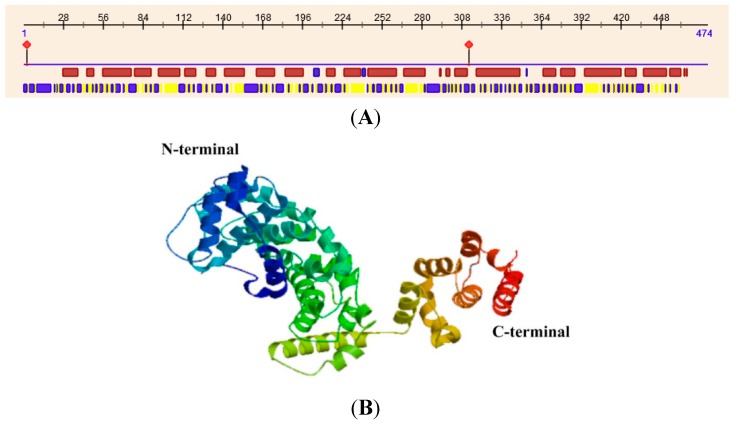
Figure 5.
Secondary and tertiary structure of MsV1H. (A) Secondary structure of MsV1H. The first line shows the secondary structure of MsV1H from RePROF prediction methods. Red boxes indicate helix, while blue boxes indicate strand. The second line shows the residues that expose to the outside or buried inside of the protein. Blue boxes and yellow boxes show exposed and buried residues, respectively. The first and 310th residues show the predicted protein binding sites; (B) Tertiary structure of MsV1H produced by Swiss-Model.

