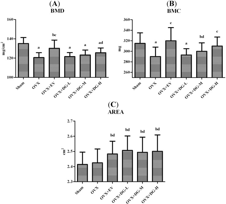
Figure 1.
Effects of diosgenin on femoral bone mineral density (BMD), bone mineral content (BMC), and projected bone area (AREA) in ovariectomy (OVX) rats. After 12-weektreatment, femurs were dissected free of soft tissue. The BMD (A); BMC (B); and AREA (C)of the femur were measured by dual-energy X-ray absorptiometry. Results are means± S.D. (n = 12 rats/group). a, p < 0.01 vs. Sham group; b, p < 0.05 vs. Sham group; c, p < 0.01 vs. OVX group; d, p < 0.05 vs. OVX group.

