Figure 3.
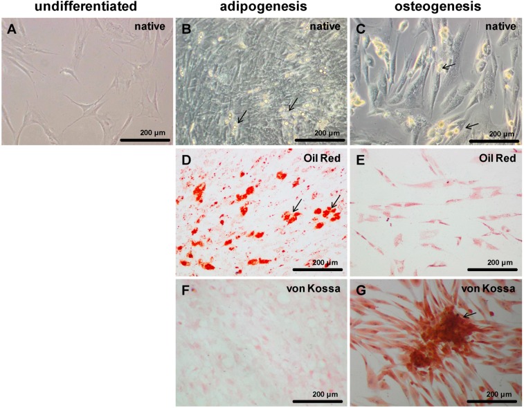
Adipogenic and osteogenic differentiated MSCs. Invert microscopical images of undifferentiated (A), adipogenic (B,D,F) and osteogenic (C,E,G) differentiated MSCs in monolayer culture. The cells were adipogenically and osteogenically differentiated for 21 days. Adipogenic differentiated cells revealed multiple fat vacuoles (B,D) which were red after oil red staining (D, arrows). Osteogenic differentiation of MSCs led to granulated elongated cells (C,E,G) which were von Kossa positive (G) and formed clusters of extracellular matrix deposits (C,G, arrows). Images of a representative experiment are shown. Scale bars = 200 µm.

