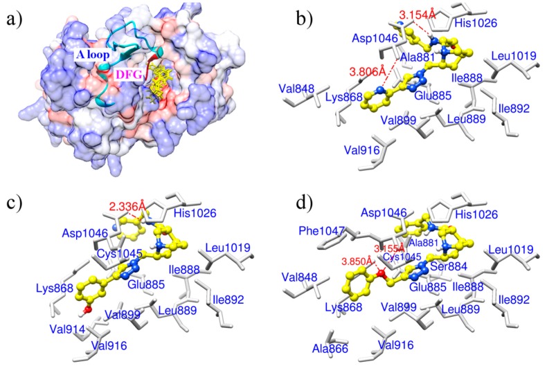
Figure 2.
(a) An overview of binding modes for the three derivatives in the binding pocket of VEGFR-2. Derivatives are shown as yellow sticks. The surface of VEGFR-2 is colored to show hydrophobicity: from dodger blue for the most hydrophilic, to white, to orange red for the most hydrophobic. The activation loop (A loop) of VEGFR-2 is displayed as cartoon in cyan and the DFG motif of the activation loop is colored in red; (b) Binding mode for ZINC08254217; (c) Binding mode for ZINC08254138; (d) Binding mode for ZINC03838680. In (b–d), sticks in light gray are related residues of VEGFR-2, hydrogen bonds labeled with distance are shown as orange dashed lines, and derivatives are in shapes of ball and stick. Color codes for atoms of derivatives in (b–d): C is yellow, N is blue, and H is light gray.

