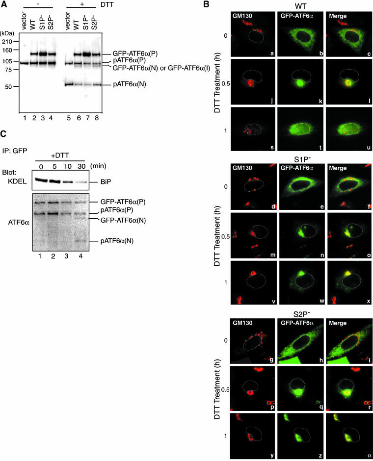
Figure 2.
Effects of mutating the S1P or S2P cleavage site on ER stress-induced processing and relocation of GFP-ATF6α. (A) Eight hours after transfection with pCMVshort-EGFP-ATF6α (WT), pC-MVshort-EGFP-ATF6α(S1P-), pCMVshort-EGFP-ATF6α(S2P-), or vector alone, CHO cells were untreated (-) or treated (+) with 1 mM DTT for 30 min before the preparation of cell lysates, which were then analyzed by immunoblotting using anti-ATF6α antibody. The migration positions of GFP-ATF6α(P), endogenous pATF6α(P), GFP-ATF6α(N), or GFP-ATF6α(I), and endogenous pATF6α(N) are marked. (B) CHO cells transfected as in A were treated with 1 mM DTT for the indicated periods, fixed, stained with anti-GM130 antibody, and then analyzed by fluorescence microscopy as described in the legend to Figure 1B. An outline of the nucleus is indicated by a white line in each cell. (C) CHO cells transfected with pCMVshort-EGFP-ATF6α were treated with 1 mM DTT for the indicated periods. GFP-ATF6α was immunoprecipitated from cell lysates using anti-GFP antibody. Immunoprecipitates were analyzed by immunoblotting using anti-ATF6α or anti-KDEL antibodies.

