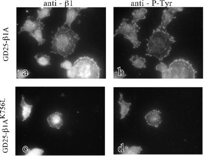
Figure 5.
Immunofluorescent detection of colocalization of β1-integrins with phosphotyrosine-containing clusters in GD25-β1A and GD25-β1AK756L cells. Double stainings for β1 (a and c) and phosphotyrosine (b and d) are shown for GD25-β1A (a and b) and GD25-β1AK756L (c and d) cells. The cells were plated on FN coated (25 μg/ml) chamber slides in the presence of the GRGDS peptide (0.2 mg/ml) and incubated at 37°C for 2 h, fixed with paraformaldehyde, and permeabilized before incubation with antibodies against β1 and phosphotyrosine.

