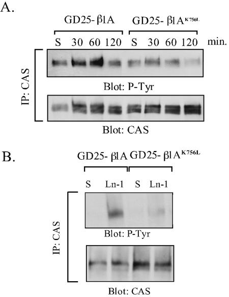
Figure 8.
Tyrosine phosphorylation of CAS in GD25-β1A and GD25-β1AK756L cells. CAS was immunoprecipitated from lysates of suspended cells (S), or cells adhering to anti-β1 mAb (A) or LN-1 (B). (A) Time curve of CAS phosphorylation after β1-mediated cell adhesion. (B) Phosphorylation of CAS in cells plated on LN-1 for 1 h. The immunoprecipitates were resolved by SDS-PAGE and transferred to nitrocellulose filters, which were blotted for phosphotyrosine (mAb RC-20) and, after stripping, for CAS protein.

