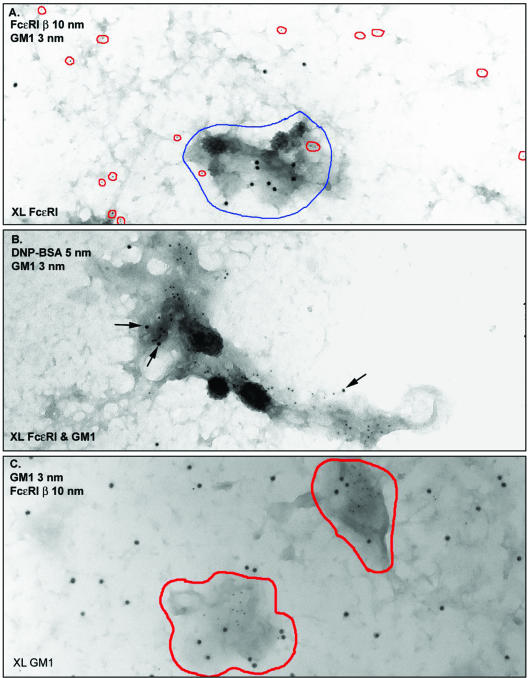
Figure 4.
GM1 and FcεRI are independently recruited to dark patches. (A) IgE-primed cells were stimulated for 2 min with DNP-BSA and then fixed and labeled with biotinyl-cholera toxin-avidin-gold (5 nm, outlined in red). Sheets were labeled with anti-FcεRI β-gold (10 nm). The electron-dense patches (outlined in blue) accumulate cross-linked FcεRI but have few particles marking GM1. (B) IgE-primed cells were incubated for 15 min at RT with biotinyl-cholera toxin, followed by 2 min at 37°C with both avidin-gold (5 nm) and DNP-BSA-gold (10 nm). Both cross-linked GM1 (abundant 5-nm gold particles) and cross-linked FcεRI (10-nm particles, arrows) are found within the same dark patch of membrane. Many small gold particles marking GM1 are found within coated vesicles. (C) GM1 was aggregated with biotinyl-cholera toxin-avidin-gold for 10 min at 37°C, membrane sheets were prepared and fixed, and then cells were labeled with antibodies marking FcεRI β. Cross-linked GM1 are exclusively found in dark patches of membrane (outlined in red), in contrast to the labeling for singlets and small clusters of uncross-linked FcεRI that are neither excluded nor concentrated in dark membrane.

