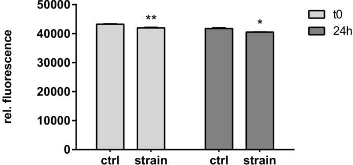
Figure 6.
Effect of strain on cell viability. C-28/I2 chondrocytes were stretched (8%) for 12 h with cyclic square waveforms at 1 Hz. Cell viability was assessed using CellTiter Blue® directly after straining (t0) or 24 h later, and is expressed as relative fluorescent signal (560 nm/590 nm ratio). Unstrained controls (ctrl), stretched cells (strain). * and ** indicate significance levels of p ≤ 0.05 and p ≤ 0.01, respectively, n = 3.

