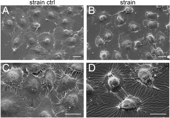
Figure 7.
Morphological changes in stretched chondrocytes. C-28/I2 cells were stretched (8%) for 12 h using cyclic square waveforms at 0.5 Hz and immediately fixed on the silicone membrane. Shown are representative scanning electron micrographs of unstrained cells (strain ctrl: A,C) and stretched cells (strain: B,D). Scale bars represent 25 μm. n = 3.

