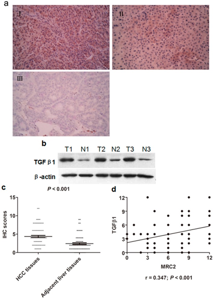
Figure 2.
(a) (I) The representative IHC staining of Transforming growth factor (TGFβ1) in HCC tissues; (II) The representative IHC staining of TGFβ1 in adjacent liver tissues; (III) The negative staining of TGFβ1 in HCC tissues; (b) The western immunoblotting results of TGFβ1 in 3 pairs of representative HCC tissues (T) and adjacent liver tissues (N); (c) The IHC scores of TGFβ1 in HCC tissues were significantly higher than one in adjacent liver tissues (p < 0.001); (d) The Spearmen rank test showed that there was positive correlation between TGFβ1 and MRC2 expression in HCC tissues (r = 0.347; p < 0.001).

