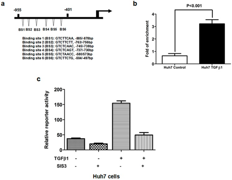
Figure 5.
(a) Schematic diagram of the potential Smad binding sites in the promoter of MRC2 which shows there are six neighboring potential Smad binding sites in the −955/−401 bp MRC2 promoter fragment; (b) With the help of ChIP assay, we obtained the DNA fragments bound with Smad protein in the nucleus of Huh7 cells. As assessed by PCR assay, TGFβ1 treatment increased significantly the occupancy of Smad protein in the MRC2 promoter; (c) The luciferase reporter assay showed that TGFβ1 treatment leaded to more MRC2 luciferase activity in Huh7 cells, which was abolished by the Smad3 inhibitor SIS3. These data strongly demonstrated that Smad3 was involved closely in the regulatory effect of TGFβ1 on MRC2 expression.

