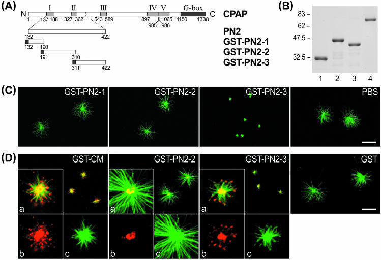
Figure 2.
Mapping the microtubule-destabilizing activity of CPAP to the PN2-3 motif. (A) Three PN2 subregions, PN2-1, PN2-2, and PN2-3, were subcloned into a pGEX-2T vector. (B) Various GST-recombinant proteins, GST-PN2-1 (lane 1), GST-PN2-2 (lane 2), and GST-PN2-3 (lane 3), were affinity purified and stained with Coomassie Blue. Lane 4 represents 1 μg of bovine serum albumin (BSA). (C and D) In vitro microtubule nucleation assay. Various GST-CPAP–truncated proteins (0.45 μM) were preincubated with purified centrosomes as described in Figure 1C. The microtubule asters were stained with FITC-conjugated anti-α-tubulin antibodies (green color) (C), or doubly stained with both anti-α-tubulin monoclonal antibodies (green) and anti-GST polyclonal antibodies (red) (D). The inserted inlet (a) in Figure 2D is a merged photograph derived from two monochrome photos, b (anti-GST antibody, red) and c (anti-α-tubulin antibody, green). Bar, 10 μm.

