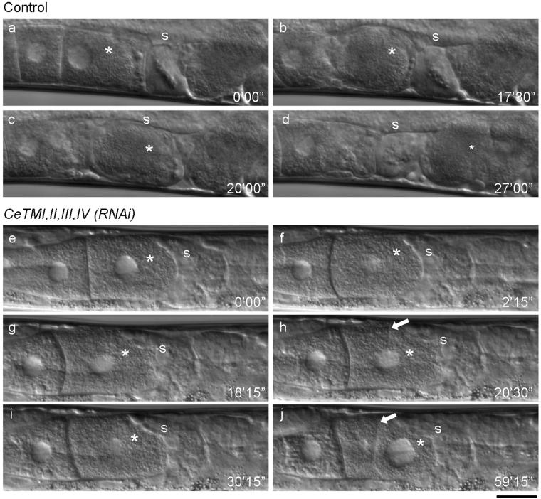
Figure 2.
Ovulation, but not oocyte maturation, is defective in the tropomyosin-RNAi worms. Ovulation processes of control (a-d) and CeTMI,II,III,IV (RNAi) (e-j) worms were recorded by time-lapse Nomarski microscopy (also see Videos 1 and 2). In a control worm, the most proximally located oocyte (a, asterisk) became mature and showed morphological change into a round shape and nuclear envelope breakdown (b). It was pushed by intense contraction of the ovary and fertilized in the spermatheca (c). Then, embryogenesis was initiated in the uterus after ovulation (d). In the CeTMI,II,III,IV (RNAi) worm, the most proximal oocyte (e, asterisk) became mature (f), but it was not ovulated due to weak contraction of the ovary (g). In the absence of ovulation, the nuclear envelope reappeared (g, asterisk) and a cleavage furrow was formed (h, arrow). The furrow was dynamic and sometimes regressed (i). However, when the cleavage is complete, the daughter cell on the left became anuclear and the large nucleus is segregated into the other cell (j). Positions of the spermatheca are indicated by “s”. Numbers indicate time (minute seconds) from the first frame. Bar, 20 μm.

