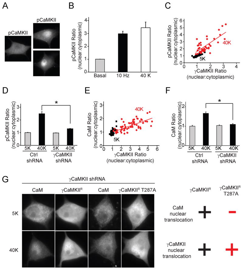
Figure 2. Activity-dependent nuclear pCaMKII redistribution is driven by γCaMKII/CaM translocation.
(A) SCG neurons were stimulated at 10 Hz for 60 s or with 40 mM K+ for 300 s and stained for phospho-Thr-286/287-CaMKII (pCaMKII). Scale bar, 10 μm. (B) Pooled data for increase in ratio of nuclear:cytoplasmic pCaMKII intensities. (C, E) Single-cell correlation of nuclear:cytoplasmic intensity ratio between pCaMKII and γCaMKII (R=0.8) or CaM and γCaMKII (R=0.7) in response to 40 mM K+, 300 s. (D, F) SCG neurons expressing γCaMKII shRNA or non-silencing control shRNA stimulated as in (C). Increased nuclear:cytoplasmic ratios for pCaMKII and CaM were prevented by γCaMKII knockdown. (G) With endogenous γCaMKII knocked down, shRNA-resistant γCaMKIIR or γCaMKIIR T287A were overexpressed in SCG neurons. Upon stimulation as in (C), both γCaMKIIR and γCaMKIIR T287A translocated to the nucleus; however, only γCaMKIIR, not γCaMKIIR T287A, rescued CaM translocation. “+” and “−“ indicate a significant or insignificant change respectively (see Supplemental Fig. 2 I, J for details). See also Figure S2.

