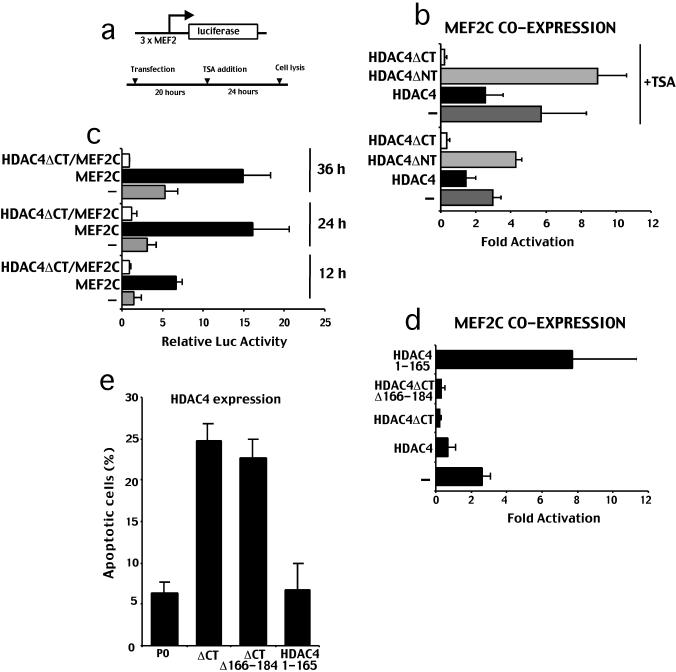
Figure 7.
HDAC4ΔC acts as a potent repressor of MEF2C-dependent transcription. (a) Schematic representation of the transfection experiments. (b) HeLa cells were transfected with 3 × -MEF2-Luc reporter (1 μg), internal control pRL-CMV(20 ng), pcDNA3.1-MEF2C (1 μg), pFLAGCMV5 (0.5 μg), and pFLAGCMV5 containing full-length HDAC4 and its deleted derivatives (0.5 μg). TSA (330 nM) was added as indicated. Data represent arithmetic means ± SD for six independent experiments. (c) HeLa cells were transfected with 3 × -MEF2-Luc reporter (1 μg), internal control pRL-CMV(20 ng), pcDNA3.1-MEF2C (1 μg), pFLAGCMV5 (0.5 μg), and pFLAGCMV5-HDAC4ΔC (0.5 μg). Cell lysates were produced at the indicated times. Expression of transfected proteins was verified using Western blotting (our unpublished data). Data represent arithmetic means ± SD for three independent experiments. (d) HeLa cells were transfected with 3 × -MEF2-Luc reporter (1 μg), internal control pRL-CMV(20 ng), pcDNA3.1-MEF2C (1 μg), pFLAGCMV5 (0.5 μg), and pFLAGCMV5 containing full-length HDAC4, HDAC4ΔC, and its deleted derivatives (0.5 μg) as indicated. Data represent arithmetic means ± SD for three independent experiments. (e) In IMR90-E1A cells pFLAGCMV5-HDAC4ΔC/Δ166-184, pFLAGCMV5-HDAC4/1-165, pFLAGCMV5-HDAC4ΔC, and pcDNA3-P0 were cotransfected together with pEGFPN1, as a reporter. The appearance of apoptotic cells was scored after 44 h from transfection. Cells showing a collapsed morphology and presenting extensive membrane blebbing were scored as apoptotic. Data represent arithmetic means ± SD for six independent experiments.

