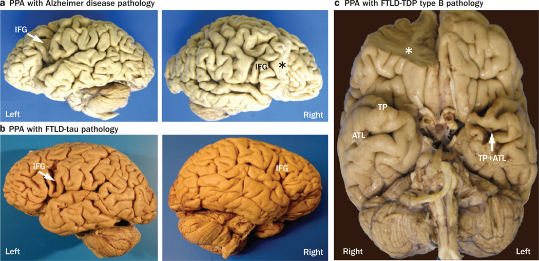
Figure 5.
Asymmetry of brain atrophy in PPA. These photographs demonstrate the asymmetric atrophy of the left-hemisphere language network associated with three different types of neuropathology in three right-handed patients with PPA.29 a | Alzheimer disease pathology in a woman who died at the age of 80 years, 7 years after the onset of PPA; the left IFG (arrow) is atrophic, but the right IFG is not. b | FTLD-tau pathology of the corticobasal degeneration type in a woman with PPA onset at the age of 72 years, who subsequently died at the age of 78 years; again, the left IFG (arrow) is atrophic, but the right IFG is not. c | Type B FTLD-TDP in a man with onset of PPA at the age of 66 years, who died 6 years later; the left TP and ATL are atrophic, but the homologous areas of the right hemisphere are not. Asterisks indicate brain regions that were removed for biochemical analyses. Abbreviations: ATL, anterior temporal lobe; FTLD-tau, frontotemporal lobar degeneration with tau protein pathology; FTLD-TDP, frontotemporal lobar degeneration with TAR DNA-binding protein 43 pathology; IFG, inferior frontal gyrus; PPA, primary progressive aphasia; TP, temporal pole. Permission obtained from Oxford University Press © Mesulam, M.-M. et al. Brain 137, 1176–1192 (2014).

