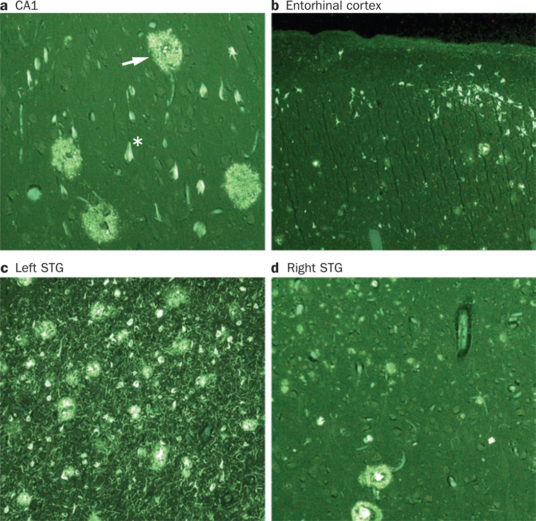
Figure 6.
Atypical distribution of AD pathology in PPA. Photomicrographs show fluorescent thioflavin-S-stained tissue from different regions of the brain of a man with PPA, with onset at age 61 years. The patient died aged 71 years and the autopsy revealed AD pathology. Plaques (arrow) and neurofibrillary tangles (asterisk) were detected in the a | CA1 area of the hippocampus, b | entorhinal cortex, c | STG of the left hemisphere and d | STG of the right hemisphere. Panels a and b show that the lesion density is lower in the CA1 and entorhinal cortex than in neocortical areas such as the STG. This pattern of lesion distribution does not conform to the principles of Braak staging in Alzheimer disease.127 Panels c and d demonstrate a higher density of plaques and neurofibrillary tangles in the STG of the left hemisphere than in the homologous region on the right side. Magnification was 100×, except in the entorhinal cortex (40× magnification). Abbreviations: AD, Alzheimer disease; PPA, primary progressive aphasia; STG, superior temporal gyrus. Permission obtained from Oxford University Press © Mesulam, M.-M. et al. Brain 137, 1176–1192 (2014).

