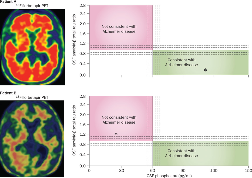
Figure 7.
Biomarker assessments to gauge the likelihood of Alzheimer pathology in PPA. Data from biomarker evaluations in two patients with a logopenic pattern of PPA at the initial clinical visit. Axial scans on the left show the results of PET imaging using an Aβ-binding tracer, 18F-florbetapir. Intense red areas on scan from Patient A are indicative of a positive PET result for Aβ plaques, indicating AD pathology, whereas the scan from Patient B demonstrates a negative result of this biomarker test. Right-hand panels show CSF levels of phospho-tau on the x-axis and a proprietary ratio of CSF Aβ:total tau on the y-axis (test performed by Athena Diagnostics). The diagnostic implications of values plotted on this graph have been determined empirically.165 Asterisks in the right-hand panels denote these values measured in CSF samples from the two patients. Patient A had high CSF levels of phospho-tau and a low CSF Aβ:total tau ratio, consistent with AD pathology, confirmed at postmortem examination. Both the CSF and PET results for Patient B are inconsistent with AD pathology and lead us to predict that the autopsy will reveal a form of frontotemporal lobar degeneration underlying PPA in this individual. Abbreviations: Aβ, amyloid-β; AD, Alzheimer disease; CSF, cerebrospinal fluid; PPA, primary progressive aphasia.

