Fig. 1.
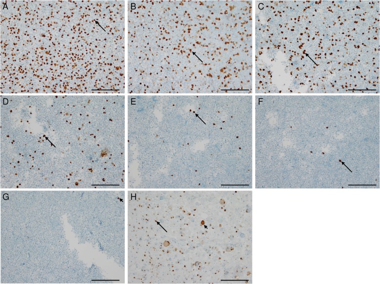
Immunocytochemistry (ICC) and immunohistochemistry (IHC) displaying detection limits in HCMV diagnostics. (A) ICC of paraffin-embedded HCMV strain Hi91-infected UKF-NB-4 neuroblastoma cells showing HCMV-IEA positive nuclei in 30%–40% of the cells (arrow). (B) ICC of paraffin-embedded HCMV-infected UKF-NB-4 cells diluted with noninfected cells in a ratio of 1:1. (C) ICC of paraffin-embedded HCMV-infected UKF-NB-4 cells diluted with noninfected cells in a ratio of 1:4. (D) ICC of paraffin-embedded HCMV-infected UKF-NB-4 cells diluted with noninfected cells in a ratio of 1:16. (E) ICC of paraffin-embedded HCMV-infected UKF-NB-4 cells diluted with noninfected cells in a ratio of 1:64. (F) ICC of paraffin-embedded HCMV-infected UKF-NB-4 cells diluted with noninfected cells in a ratio of 1:256. (G) ICC of paraffin-embedded HCMV-infected UKF-NB-4 cells diluted with noninfected cells in a ratio of 1:1024. (H) Cerebellar tissue from a patient with HCMV encephalitis showing atypical HCMV-positive nuclei (arrow) of infected neurons and positive nuclei of surrounding glial cells (arrow-heads). (Scale 100 µm).

