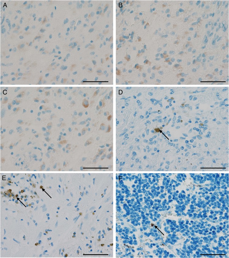
Fig. 4.
Old intratumoral bleeding and fixation artefacts are potential pitfalls in immunohistochemistry-based HCMV diagnostics. (A) Positive glioblastoma case with gemistocytic cells being positive for HCMV-pp65 antigen and also for (B) HCMV immediate early antigen (IEA). (C) Same glioblastoma case stained without primary antibody also showing a similar positive signal indicating an unspecific reaction. (D and E) Neuroblastoma with hematoidin artefacts appearing like positive tumor cells (arrows). (F) Neuroblastoma with formalin pigments (arrow). (Scale 50 µm).

