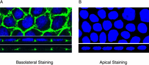
Figure 2.
Localization of endogenous type I TGFβ receptors in polarized MDCK monolayers. MDCK cells were plated in 12-mm Transwells and allowed to polarize >72 h. Receptors were imaged upon selective basolateral (A) or apical (B) immunohistochemical staining with an endogenous type I TGFβ receptor primary rabbit antibody coupled to an antirabbit Alexa 488 secondary antibody (green). XY (horizontal) sections are in the top image and XZ (vertical) sections are shown in the lower image. Nuclei were stained with DAPI.

