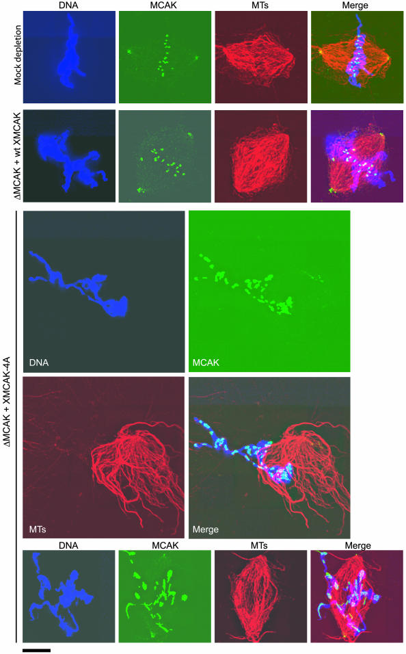
Figure 6.
XMCAK-4A fails to localize to inner centromeres. Structures assembled in mock-depleted extract, and MCAK-depleted extracts (ΔMCAK) that were supplemented with either wt XMCAK or XMCAK-4A were fixed and stained with anti-XMCAK antibodies (green). Tubulin (MTs; red) was visualized with rhodamine-tubulin and DNA (blue) with Hoechst 33342. Bar, 10 μM.

