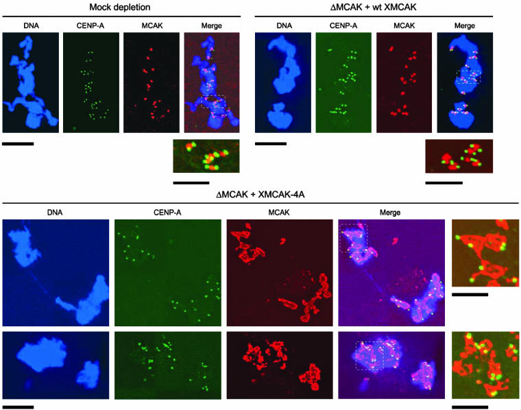
Figure 7.
XMCAK-4A does not exhibit preferential localization to centromeres. Structures assembled in mock-depleted extract, and MCAK-depleted extracts (ΔMCAK) supplemented with either wt XMCAK or XMCAK-4A were fixed and double stained with antibodies specific for XMCAK (red) and CENP-A (green). DNA (blue) was visualized with Hoechst 33342. The boxed regions are enlarged and shown without DNA staining to allow more careful comparison of XMCAK and CENP-A immunofluorescence. Bars, 10 μM except for the enlarged images, which are 5 μM.

