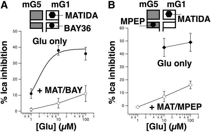
Fig. 5.
Effect of combined inhibition by MATIDA and selective NAMs on the partial glutamate concentration response of mGluR1/mGluR5. (A) Effect of glutamate (1–100 µM) applied alone (black circles) in SCG neurons expressing mGluR1 and mGluR5, and in the presence of 100 µM MATIDA and 1 µM BAY36 (open circles). All drug applications were paired in the same cells (n = 6 for all data points). (B) Effect of glutamate (10–100 µM) applied alone (black diamonds) in SCG neurons expressing mGluR1 and mGluR5, and 1–100 µM glutamate in the presence of 100 µM MATIDA and 1 µM BAY36 (open diamonds). All drug applications were paired in the same cells (n = 3 for all data points). BAY, BAY36; MAT, MATIDA.

