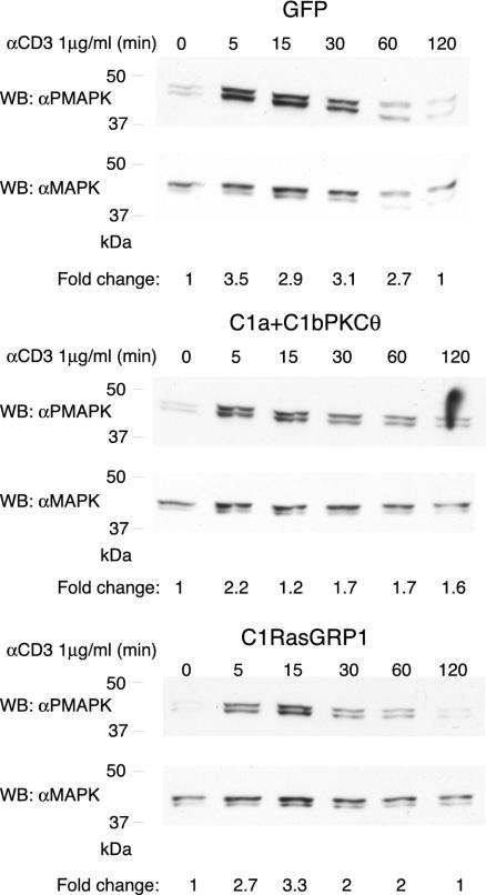
Figure 8.
C1RasGRP and C1a+C1bPKCθ modify Ras activation in response to soluble CD3 activation. Cells transfected with GFP, C1RasGRP or C1a+C1bPKCθ were sorted to recover GFP-positive cells. GFP expression was assessed by flow cytometry after 8 h culture; cells were then serum-starved (1 h), stimulated with soluble CD3 (1 μg/ml) for the times indicated, then collected and processed (see MATERIALS AND METHODS) for Western blot. Analysis with PMAPK antibody showed Jurkat cell activation differences according to the construct transfected. Anti-MAPK antibody was used as a protein loading control. Results are representative of four independent experiments. GFP and GFPC1RasGRP cells were 95% GFP-positive; GFPC1a+C1bPKCθ cells were 78% GFP-positive. The x-fold change in activation for each condition was estimated by densitometric analysis of filters. Values (arbitrary units) were normalized taking into consideration the protein levels of each sample as determined by Western blot analysis.

