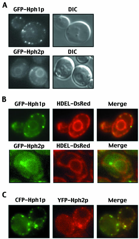
FIG. 6.
Hph1p and Hph2p localize to the ER. (A) Wild-type yeast cells (YPH499) expressing GFP-Hph1p (pVH1) or GFP-Hph2p (pVH2) under a methionine-repressible promoter were grown to log phase and visualized by fluorescence microscopy and differential interference microscopy (DIC) (see Materials and Methods). (B) GFP-Hph1p (pVH1) or GFP-Hph2p (pVH2) was expressed in wild-type yeast cells expressing HDEL-DsRed (VHY87) to mark the ER. In the third panel, the two images were merged, and yellow regions indicate areas of overlapping localization. (C) Wild-type cells (YPH499) were transformed with plasmids to express CFP-Hph1p (pVH10) and YFP-Hph2p (pVH11) and visualized by fluorescence microscopy. In the third panel, the two images were merged, and yellow regions indicate areas of overlapping localization.

