Fig. 5.

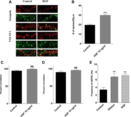
Effect of HGF treatment on synaptogenesis in dissociated hippocampal neurons. HGF treatment supports the formation of functional synapses as indicated by a high correlation between postsynaptic spines (red) and markers of presynaptic active zones (green). (A) Representative images of hippocampal neurons transfected with mRFP-β-actin on DIV6 and treated with 10 ng/ml of HGF or vehicle for 5 days. The neurons were stained for the general presynaptic marker synapsin and the glutamatergic presynaptic marker VGLUT1. (B) Bar graph demonstrating an active phenotype as indicated by a significant increase in the number of spines per 50 μm of dendrite length following stimulation with HGF (10 ng/ml). Mean number of spines, HGF = 33 versus control = 23; mean ± S.E.M.; N = 25; ***P < 0.001. (C) Percent correlation of actin-enriched postsynaptic spines (red) juxtaposed to the universal presynaptic marker synapsin (green). NS = P > 0.05. The high percent correlation suggests functional synapses are formed. (D) Percent correlation of actin-enriched spines (red) juxtaposed to the glutamatergic presynaptic marker VGLUT1 (green). NS = P > 0.05. A greater than 95% correlation suggests many of these inputs are glutamatergic. (E) Effect of dihexa and HGF treatment on the frequency of mEPSCs in dissociated hippocampal neurons. Dissociated hippocampal neurons transfected with mRFP-β-actin were stimulated with 10–12 M dihexa or 10 ng/ml for 5 days prior to recording mEPSCs. Neurons were treated with tetrodotoxin, picrotoxin, and strychnine to block sodium channels, GABAA, and glycine receptors. Treatment with both agonists significantly enhanced AMPA-receptor mediated currents compared to vehicle treated neurons Mean ± S.E.M.; n = 9 respectively; ***P < 0.012 HGF versus control; P < 0.04 Hex versus control). NS, not significant.
