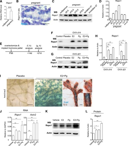
Figure 3.
Hormones induce the expression of Rspo1 in vivo and in vitro. (A) Rspo1 qPCR analysis indicated that Rspo1 expression was significantly increased in late pregnancy mammary epithelial cells. (***) P < 0.01. (B) In situ hybridization showed the robust Rspo1 level in pregnancy (in cyan). Nuclei were counterstained with hematoxylin (in purple). Bar, 20 μm. (C,D) Western blot and quantification indicate the Rspo1 expression during pregnancy, lactation, and involution. (***) P < 0.01. (E) Mice were ovariectomized and implanted with E2 or Pg pellets alone or in combination (E2 + Pg) for 14 or 21 d. (F–H) Mammary glands of control and ovariectomized mice (OVX) were harvested and analyzed by Western blot and quantification. Either E2 or Pg treatment was sufficient to enhance Rspo1 expression, while the augmentation was more pronounced in the presence of both E2 and Pg. The estrous cycle was not controlled in these experiments. (***) P < 0.01. (I) Axin2-lacZ mice were ovariectomized and implanted with E2 and Pg pellets for 21 d. The mammary glands were harvested and subjected to X-gal and K14 staining. Axin2 expression was induced in both basal cells (arrow in insert 1) and luminal cells (arrowhead in insert 2) by hormonal treatment. Bars: in the whole mount, 1 mm; in sections, 20 μm. (J) qPCR analysis of cultured luminal cells indicated that Rspo1 and Axin2 expression were significantly increased in all conditions of hormonal treatment. (***) P < 0.01. (K,L) Western blot analysis of cultured luminal cells indicated that the Rspo1 level significantly increased in the presence of hormones.(***) P < 0.01; (*) P < 0.05.

