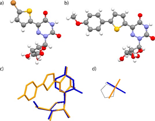
Figure 2.

(a, b) X-ray crystal structures of 4 and 1d, respectively; (c) overlay of X-ray the crystal structure of 1c (orange) with uridine (blue); overlaying the ribose rings shows minimal impact on the sugar pucker (rmsd = 0.04 Å); (d) schematic top view illustrating the relative conformation of the nucleobases in uridine (blue) and 1c (orange).
