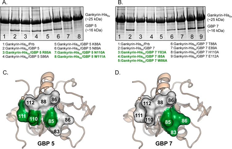
Figure 3.
(A) Coomassie-stained PAGE following copurification of gankyrin-His6x and untagged Prb, gankyrin binding protein 5 (GBP 5), and alanine mutants thereof (stated below the gel). (B) Coomassie-stained PAGE following copurification of gankyrin-His6x and untagged Prb, gankyrin binding protein 7 (GBP 7), or alanine mutants thereof (stated below the gel). (C) Binding face of GBP 5, with key gankyrin-binding residues highlighted in green. (D) Binding face of GBP 7, with key gankyrin-binding residues highlighted in green. Structures shown in panels C and D are of the putative binding face of Prb, which is the starting point for our protein resurfacing. These representations are not intended to provide any information on structural features of GBP 5 or GBP 7, or alanine mutants thereof, but rather to graphically represent where mutations deleterious to gankyrin binding reside on GBP 5 and GBP 7. Taken together, these depictions indicate where binding “hot spots” are on the resurfaced proteins GBP 5 and GBP 7, as determined by our pull-down data in panels A and B.

