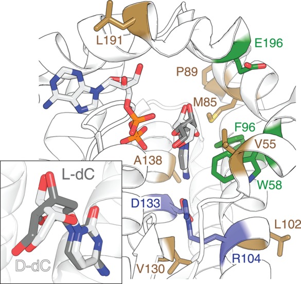
Figure 2.

Summary of amino acid substitution in human dCK with bound ADP in the phosphoryl donor site, as well as d- and l-dC in the phosphoryl acceptor binding pocket (PDB access codes: 2NO1 and 2NO7(30)). Variant positions R104M and D133N in ssTK3 are marked in violet. The three positions probed in Library A are highlighted in green, while the seven residues varied in Library B are colored in brown. Insert: Overlay of the d- and l-dC bound in the active site shows the highly similar positioning of the pyrimidine moiety, as well as the 3′- and 5′-hydroxyl groups.
